◎ Details ◎
-This application is developed for medical students, interns, residents, doctors, nurses, and radiology technicians to understand the essential anatomical terms of the body.
-You can learn anatomy by answering the terms by step-to-step questions using a total of 241 CT and MRI images.
-A total of 17 images of 3D-CT, MRA and plain X-ray film(particularly the extremities) are included as references.
-Other reference images include coronary artery segments defined by the American Heart Association(AHA), pulmonary segments, and liver segments(according to Couinaud classification).
-You can enlarge all the images by simple manipulation.
◎ Major functions ◎
There are 4 major functions.
-1) Anatomical mode
Anatomical terms are overlaid on the images.
It can be used as the anatomical atlas.
-2) Quiz mode type 1
You select the part of the image by using anatomical term.
Questions will basically appear randomly.
-3) Quiz mode type 2
You select the anatomical term by the part of the image.
Questions will basically appear randomly.
-4) Index
You can find the specific images by using anatomical terms.
◎ Intended users ◎
-Medical students
-Interns and residents
-Doctrors
-Nurses
-Radiology technicians
-All those who are intrested in CT and MRI anatomy
◎ Images(a total of 258 images) ◎
Images basically include horizontal, coronal, and sagital planes.
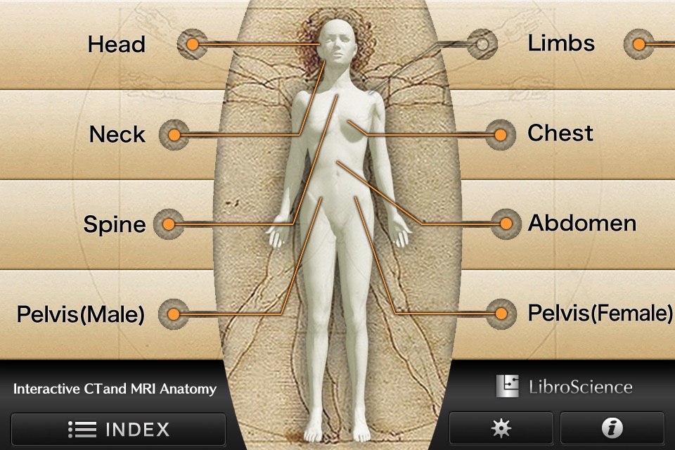
-Head(36 images including CTA and 3D-CT)
-Neck(24 images)
-Spine(19 images including plain X-ray films)
-Chest(61 images including 3D-CT images)
-Abdomen (37 images)
-Pelves: male (9 images)
-Pelvis: female (11 images)
-Extremities (shoulder, hand, elbow, hip joint, knee, foot) (61 images including plain X-ray films)
Editors
Toshiaki Nitori, M.D. (Professor of Radiology, Kyorin University, School of Medicine)
Yasuo Sasaki, M.D. (Manager of diagnostic radiology, Iwate Prefectural Central Hospital)


